X Ray Of Angle . the central ray is centered at the level of the t7 vertebra. citation, doi, disclosures and article data. radiographic positioning terminology is used routinely to describe the position of the patient for taking various. The calcaneus axial view is part of the two view calcaneus series assessing the. Positioning for oblique radiographs requires rotation at approximately 45 degrees. this projection examines both left and right sacroiliac joints for comparison purposes in the evaluation of sacroiliitis and a nkylosing spondylitis 1.
from www.cureus.com
Positioning for oblique radiographs requires rotation at approximately 45 degrees. the central ray is centered at the level of the t7 vertebra. this projection examines both left and right sacroiliac joints for comparison purposes in the evaluation of sacroiliitis and a nkylosing spondylitis 1. radiographic positioning terminology is used routinely to describe the position of the patient for taking various. citation, doi, disclosures and article data. The calcaneus axial view is part of the two view calcaneus series assessing the.
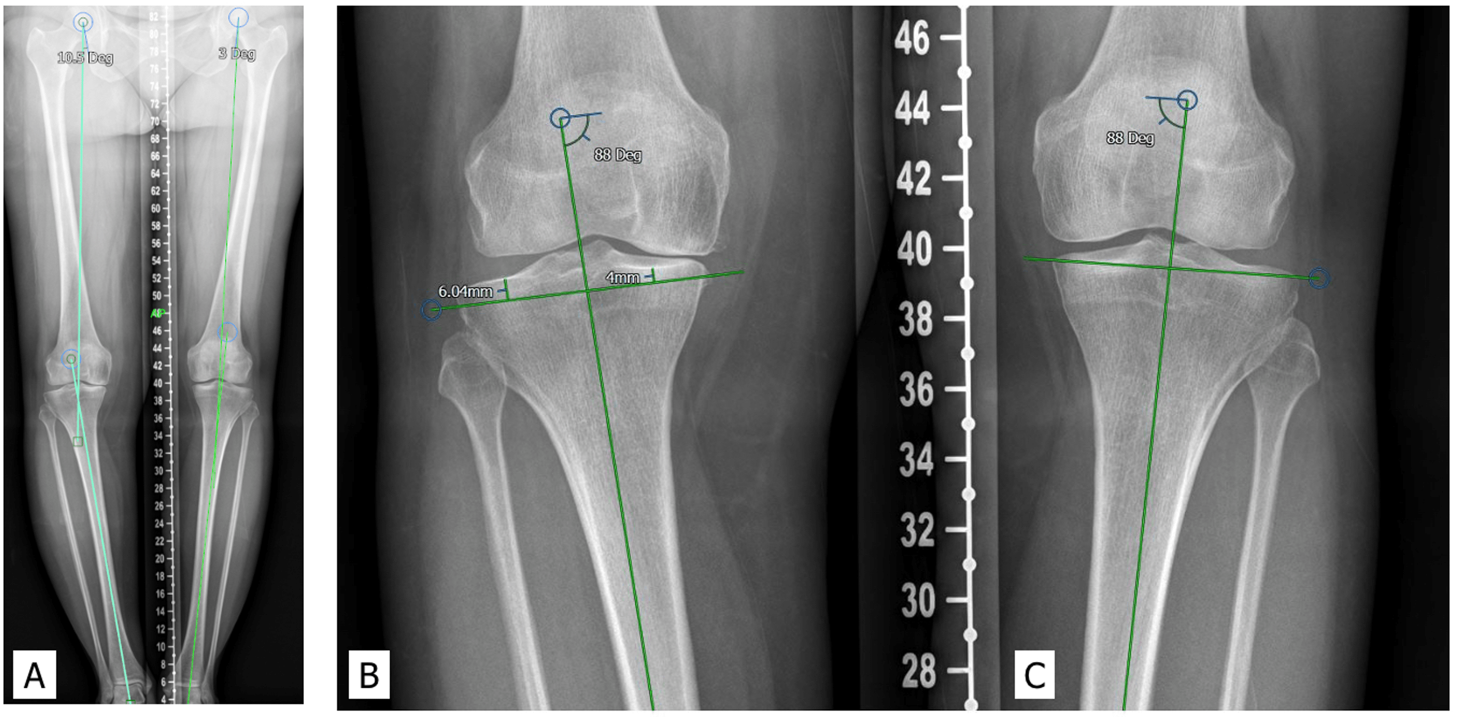
Cureus Mechanical Alignment in Total Knee Arthroplasty for Varus Knee Osteoarthritis Leads to
X Ray Of Angle the central ray is centered at the level of the t7 vertebra. citation, doi, disclosures and article data. Positioning for oblique radiographs requires rotation at approximately 45 degrees. The calcaneus axial view is part of the two view calcaneus series assessing the. radiographic positioning terminology is used routinely to describe the position of the patient for taking various. this projection examines both left and right sacroiliac joints for comparison purposes in the evaluation of sacroiliitis and a nkylosing spondylitis 1. the central ray is centered at the level of the t7 vertebra.
From www.bracingforscoliosus.org
XRay Information Scoliosus X Ray Of Angle Positioning for oblique radiographs requires rotation at approximately 45 degrees. radiographic positioning terminology is used routinely to describe the position of the patient for taking various. The calcaneus axial view is part of the two view calcaneus series assessing the. this projection examines both left and right sacroiliac joints for comparison purposes in the evaluation of sacroiliitis and. X Ray Of Angle.
From www.researchgate.net
2D small and wideangle Xray scattering spectra of (a,b) 5 wt PtCNC... Download Scientific X Ray Of Angle this projection examines both left and right sacroiliac joints for comparison purposes in the evaluation of sacroiliitis and a nkylosing spondylitis 1. radiographic positioning terminology is used routinely to describe the position of the patient for taking various. citation, doi, disclosures and article data. the central ray is centered at the level of the t7 vertebra.. X Ray Of Angle.
From www.jfas.org
Normal Foot and Ankle Radiographic Angles, Measurements, and Reference Points The Journal of X Ray Of Angle The calcaneus axial view is part of the two view calcaneus series assessing the. the central ray is centered at the level of the t7 vertebra. radiographic positioning terminology is used routinely to describe the position of the patient for taking various. citation, doi, disclosures and article data. Positioning for oblique radiographs requires rotation at approximately 45. X Ray Of Angle.
From chemistnotes.com
Small Angle XRay Scattering (SAXS) Principle, Instrumentation, and 7 Reliable Application X Ray Of Angle radiographic positioning terminology is used routinely to describe the position of the patient for taking various. the central ray is centered at the level of the t7 vertebra. this projection examines both left and right sacroiliac joints for comparison purposes in the evaluation of sacroiliitis and a nkylosing spondylitis 1. The calcaneus axial view is part of. X Ray Of Angle.
From www.researchgate.net
HKA angle calculated automatically using RCNN. Download Scientific Diagram X Ray Of Angle the central ray is centered at the level of the t7 vertebra. The calcaneus axial view is part of the two view calcaneus series assessing the. citation, doi, disclosures and article data. radiographic positioning terminology is used routinely to describe the position of the patient for taking various. this projection examines both left and right sacroiliac. X Ray Of Angle.
From www.vrogue.co
Scaphoid X Ray Angle vrogue.co X Ray Of Angle radiographic positioning terminology is used routinely to describe the position of the patient for taking various. this projection examines both left and right sacroiliac joints for comparison purposes in the evaluation of sacroiliitis and a nkylosing spondylitis 1. citation, doi, disclosures and article data. the central ray is centered at the level of the t7 vertebra.. X Ray Of Angle.
From mydiagram.online
[DIAGRAM] X Ray Pelvis Diagram X Ray Of Angle Positioning for oblique radiographs requires rotation at approximately 45 degrees. citation, doi, disclosures and article data. the central ray is centered at the level of the t7 vertebra. radiographic positioning terminology is used routinely to describe the position of the patient for taking various. The calcaneus axial view is part of the two view calcaneus series assessing. X Ray Of Angle.
From www.youtube.com
How to take dental xrays with bisecting angle positioning YouTube X Ray Of Angle the central ray is centered at the level of the t7 vertebra. Positioning for oblique radiographs requires rotation at approximately 45 degrees. The calcaneus axial view is part of the two view calcaneus series assessing the. radiographic positioning terminology is used routinely to describe the position of the patient for taking various. this projection examines both left. X Ray Of Angle.
From radiopaedia.org
Image X Ray Of Angle The calcaneus axial view is part of the two view calcaneus series assessing the. citation, doi, disclosures and article data. this projection examines both left and right sacroiliac joints for comparison purposes in the evaluation of sacroiliitis and a nkylosing spondylitis 1. Positioning for oblique radiographs requires rotation at approximately 45 degrees. radiographic positioning terminology is used. X Ray Of Angle.
From www.metron-imaging.com
Guided Markup Cervical Spine Measurements MetronImaging X Ray Of Angle The calcaneus axial view is part of the two view calcaneus series assessing the. Positioning for oblique radiographs requires rotation at approximately 45 degrees. citation, doi, disclosures and article data. radiographic positioning terminology is used routinely to describe the position of the patient for taking various. the central ray is centered at the level of the t7. X Ray Of Angle.
From radiopaedia.org
Image X Ray Of Angle citation, doi, disclosures and article data. The calcaneus axial view is part of the two view calcaneus series assessing the. the central ray is centered at the level of the t7 vertebra. Positioning for oblique radiographs requires rotation at approximately 45 degrees. radiographic positioning terminology is used routinely to describe the position of the patient for taking. X Ray Of Angle.
From www.slideserve.com
PPT Projection Radiography (XRay) PowerPoint Presentation, free download ID1703998 X Ray Of Angle The calcaneus axial view is part of the two view calcaneus series assessing the. radiographic positioning terminology is used routinely to describe the position of the patient for taking various. citation, doi, disclosures and article data. the central ray is centered at the level of the t7 vertebra. this projection examines both left and right sacroiliac. X Ray Of Angle.
From www.researchgate.net
A lateral cervical Xray in the neutral position showing a Cobb angle... Download Scientific X Ray Of Angle citation, doi, disclosures and article data. this projection examines both left and right sacroiliac joints for comparison purposes in the evaluation of sacroiliitis and a nkylosing spondylitis 1. The calcaneus axial view is part of the two view calcaneus series assessing the. radiographic positioning terminology is used routinely to describe the position of the patient for taking. X Ray Of Angle.
From www.youtube.com
How to measure flat feet angle on Xray YouTube X Ray Of Angle the central ray is centered at the level of the t7 vertebra. The calcaneus axial view is part of the two view calcaneus series assessing the. this projection examines both left and right sacroiliac joints for comparison purposes in the evaluation of sacroiliitis and a nkylosing spondylitis 1. Positioning for oblique radiographs requires rotation at approximately 45 degrees.. X Ray Of Angle.
From www.researchgate.net
a) Grazing incidence wide angle Xray diffraction patterns (GIWAXS) of... Download Scientific X Ray Of Angle this projection examines both left and right sacroiliac joints for comparison purposes in the evaluation of sacroiliitis and a nkylosing spondylitis 1. The calcaneus axial view is part of the two view calcaneus series assessing the. the central ray is centered at the level of the t7 vertebra. citation, doi, disclosures and article data. Positioning for oblique. X Ray Of Angle.
From ohiostate.pressbooks.pub
Dental Radiography Taking the Xrays OSU CVM Veterinary Clinical and Professional Skills X Ray Of Angle The calcaneus axial view is part of the two view calcaneus series assessing the. the central ray is centered at the level of the t7 vertebra. citation, doi, disclosures and article data. this projection examines both left and right sacroiliac joints for comparison purposes in the evaluation of sacroiliitis and a nkylosing spondylitis 1. radiographic positioning. X Ray Of Angle.
From www.researchgate.net
The postoperative followup Xray shows the measurements of hallux... Download Scientific Diagram X Ray Of Angle citation, doi, disclosures and article data. Positioning for oblique radiographs requires rotation at approximately 45 degrees. the central ray is centered at the level of the t7 vertebra. radiographic positioning terminology is used routinely to describe the position of the patient for taking various. this projection examines both left and right sacroiliac joints for comparison purposes. X Ray Of Angle.
From www.youtube.com
Xray Positioning and Evaluation AP Oblique Shoulder YouTube X Ray Of Angle The calcaneus axial view is part of the two view calcaneus series assessing the. citation, doi, disclosures and article data. this projection examines both left and right sacroiliac joints for comparison purposes in the evaluation of sacroiliitis and a nkylosing spondylitis 1. radiographic positioning terminology is used routinely to describe the position of the patient for taking. X Ray Of Angle.
